


CPTCast Slide Image Index - Click on the images below to enlarge

BMT Scheme
Here’s a slide from the BMT scheme. Marks were deducted for background staining and non specific silver deposit which are masking the demonstration of the fibres. This slide scored 5/10.

NADH - Muscle Scheme
Here’s an example of a slide from our Muscle Scheme showing the NADH method. This image shows excellent fibre type demonstration and scored 10/10 at assessment!
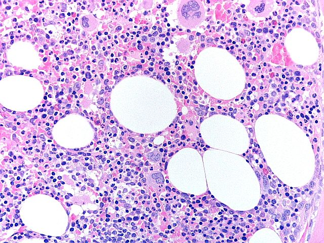
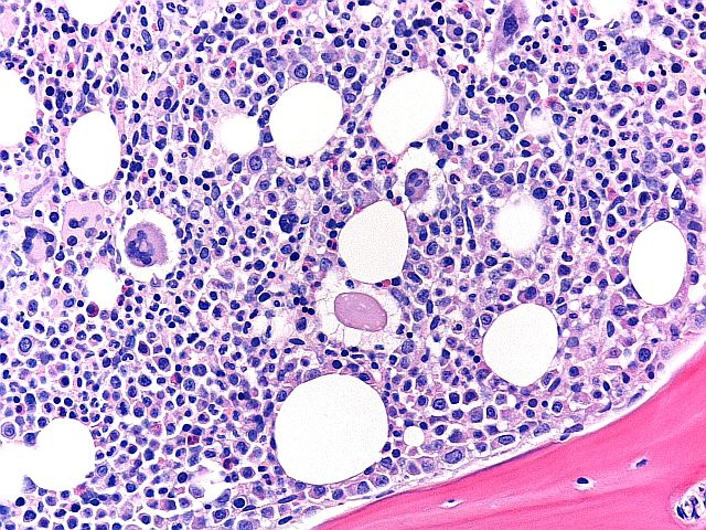
H&E - BMT Scheme
This is an excellent preparation of a BMT biopsy, with balanced H&E staining, good chromatin detail, and selective staining of cell types within the bone marrow tissue. This slide was scored 9/10 at assessment.
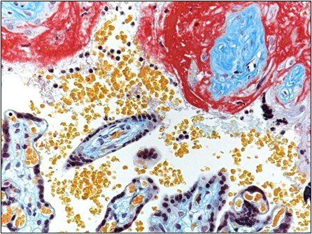
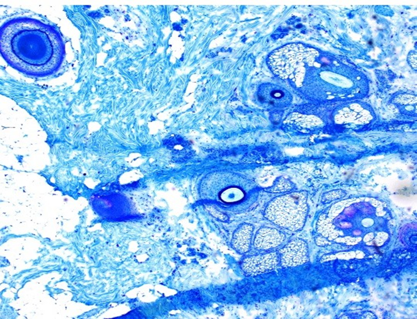
Martius Scarlet Blue
This is an example of a slide stained by Martius Scarlet Blue to demonstrate Fibrin.
All the fibrin should be brightly coloured and distinguishable from other structures.There should be good colour separation between the dyes for red blood cells (yellow), muscle (red) and collagen (blue). The nuclear staining should be dark blue or black
This is an excellent example that scored 10/10.

PAS - Renal Scheme
Seen at x20, this is an example of a PAS stain.Marks were deducted for ‘primary background staining.’ Although the glomerular basement membranes are well stained, the contrast could be better if there was less background Schiff staining. In addition, water droplets were seen under the coverslip.
The overall score for this section scored 7/10.
Gomori’s Trichrome – Fresh Muscle
This is example of a poorly executed preparation followed by poor staining. As is with all fresh muscle staining methods, good preperation is key. If your lab is having difficulties you can access Best Methods Protocols and additional quality control assessments.
This scored 2/10.
Papanicolaou - Diagnostic Cytology Scheme
This is an example of a Body Cavity fluid stained by Papanicolaou. The preparation thickness is uneven. The Nuclear stain is too intense, masking cytoplasmic stain and there is indistinct chromatin detail. This slide score 6/10.

Glail Fibres - Neuropathology Scheme
Seen at x20, this is an example of a technique for th demonstration of glail fibres. The staining seen here is suboptimal with weak fibres. This scored 7/10.
H+E - MOHS Scheme
This image shows an H+E stained Mohs frozen section, seen at x4, demonstrating a beautiful pancake technique. This slide scored top marks of 10/10.
Mega Block H+E- Mega Block companion scheme
This slide shows significant nuclear bubbling and nuclear meltdown. These artefacts suggest incomplete fixation and under or incorrect processing.
This slide scored 5/10.
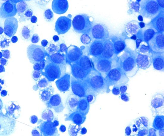
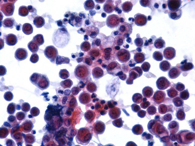
Fluid Romanowsky - Diagnostic Cytology Scheme.
This slide shows excellent staining of both nucleus and cytoplasm.
This scored 9/10 at assessment.
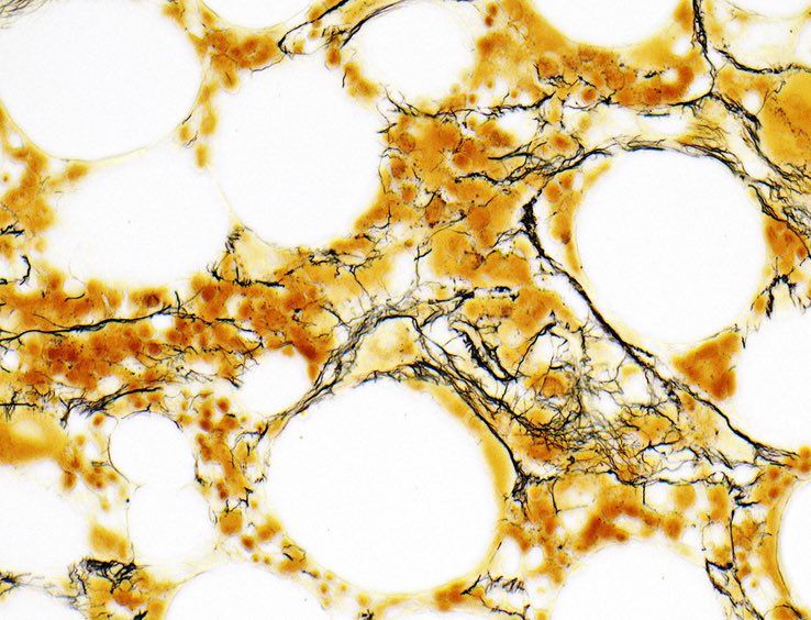
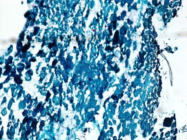
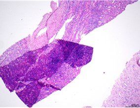
Reticulin Staining - BMT Scheme
This is an excellent example of retuculin staining. The silver has impregnated course and fine fibre networks with high specificity.
This scored 9/10.
Toludine Blue - Mohs Scheme
This section of hair bearing skin demonstrates adequate staining but the presence of two clear folds in the section were noted. This slide scored 7/10.

Masson Fontanta - Specialist Techniques Scheme
Although this counterstain may look a little heavy on this photomicrograph image, when comparing this section to others in the run it was felt an excellent score was justified as it fulfilled all the assessment criteria in the Specialist Techniques. This slide scored 10/10.

H+E - Mega Block Scheme
There is significant morphological damage with loss of glandular architecture; caused by poor fixation and processing. Megablocks generally require extended or enanced fixation and processing schedules. This slide score a low 2/10.

H+E - Mohs Scheme
This H+E stained slide shows folds and creases, and freezing artefact. There are holes and trimming artefact and the full epidermis is not visible. The Haematoxylin intensity is too weak. This slide scored 4/10.
H+E - BMT Scheme
Seen at x20. Overall a very good preperation, but marks were deducted for folds and creases, loss of material, and haematoxylin background staining. This slide scored 8/10.

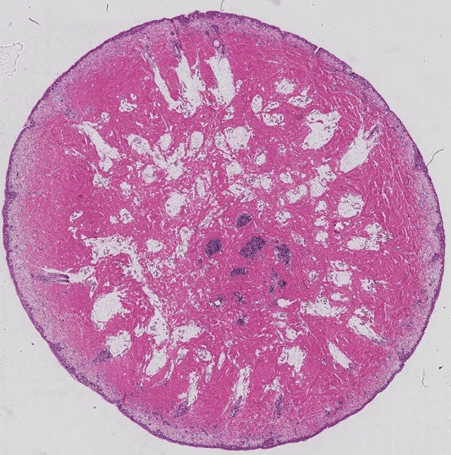
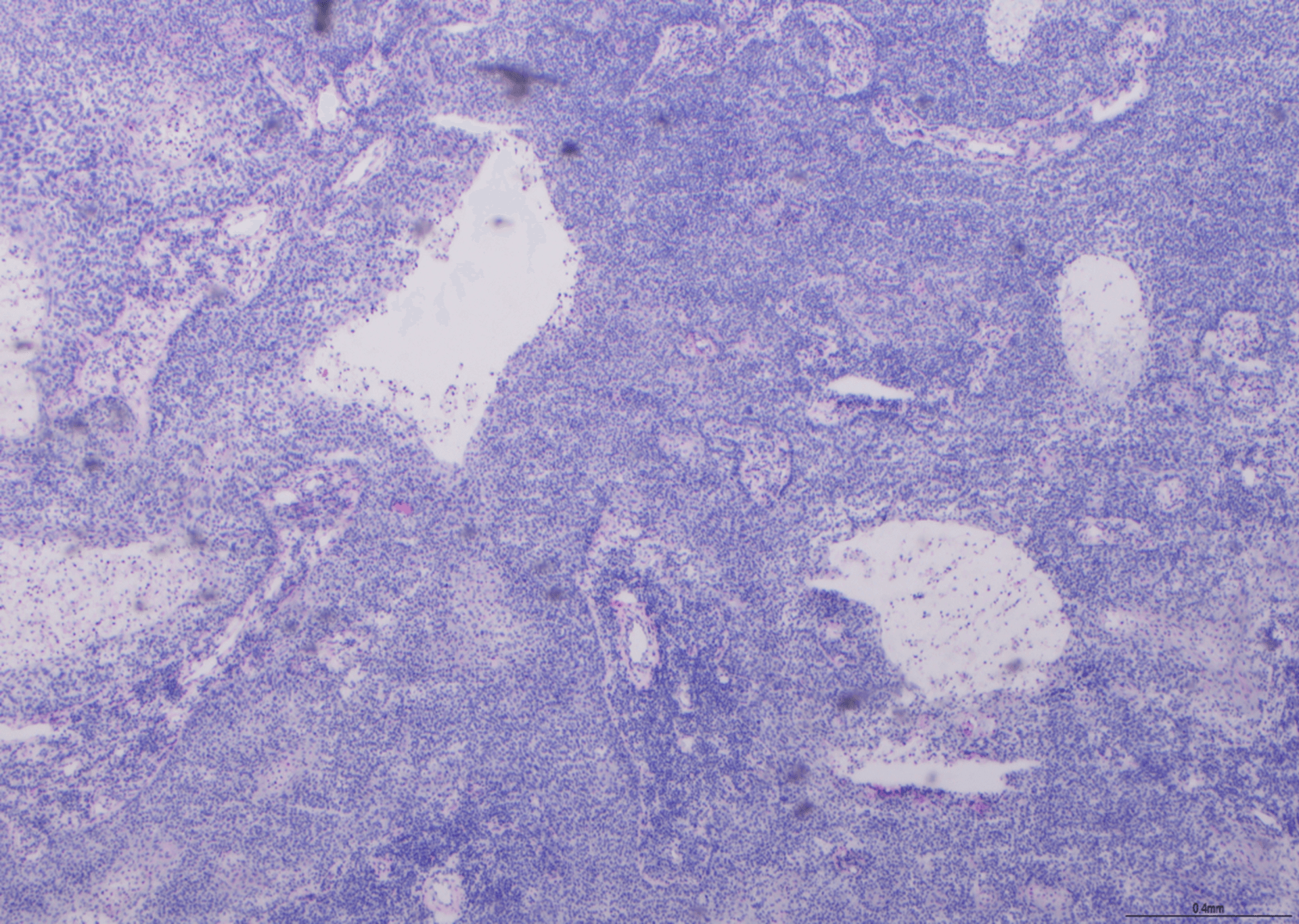
Methenamine Silver - Renal Scheme
Seen at 10x magnification. There were no glomeruli present, which makes the assessment of glomerular basement membrane staining impossible. The level of silver impregnation was also considered too strong, as seen in the tubules here. Marks were deducted as this section was considered 'insufficient for assessment'. This slide scored 2/10.
Glial Fibre - Neuropathology Scheme
Seen at x4, this slide shows very weak demonstration. This slide scored 4/10.

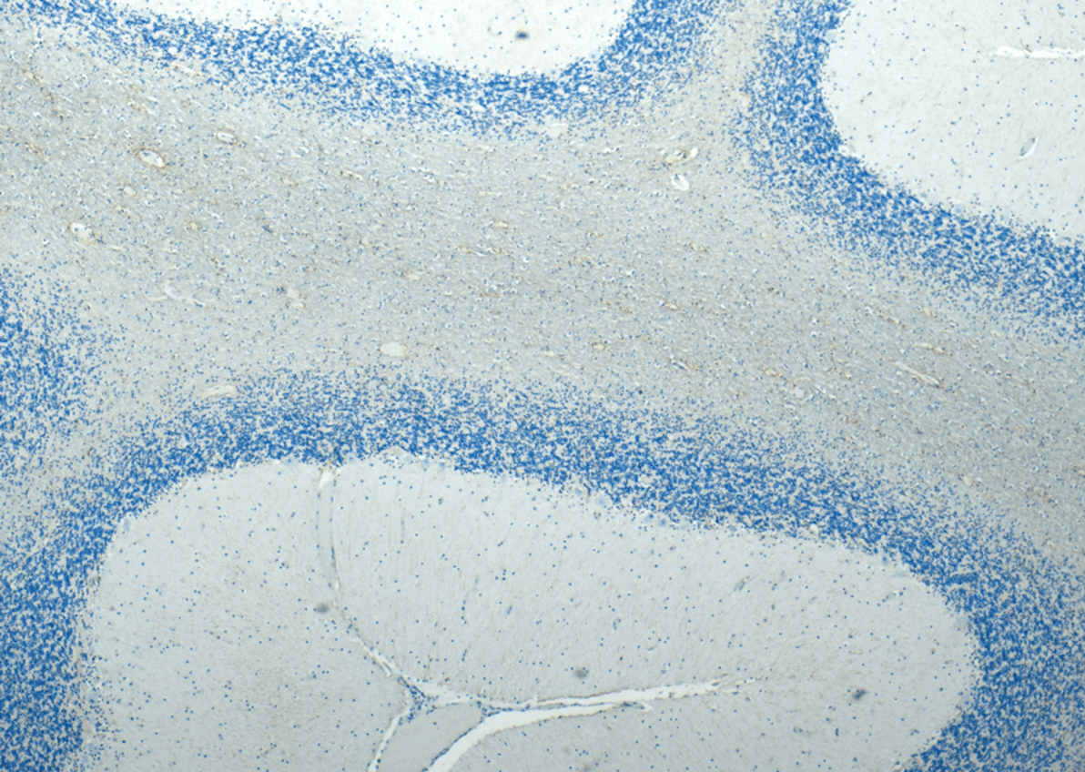
Glial Fibre - Neuropathology Scheme
Seen at x4, this slide shows excellent demonstration. This slide scored 10/10.

Acid Phosphatase - Muscle Scheme
Seen at x10, this slide is an example of a poorly stained slide. Marks were deducted for air bubbles, lifitng and non selectivity of staining as well as primary stain deposit. This slide scored 2/10.
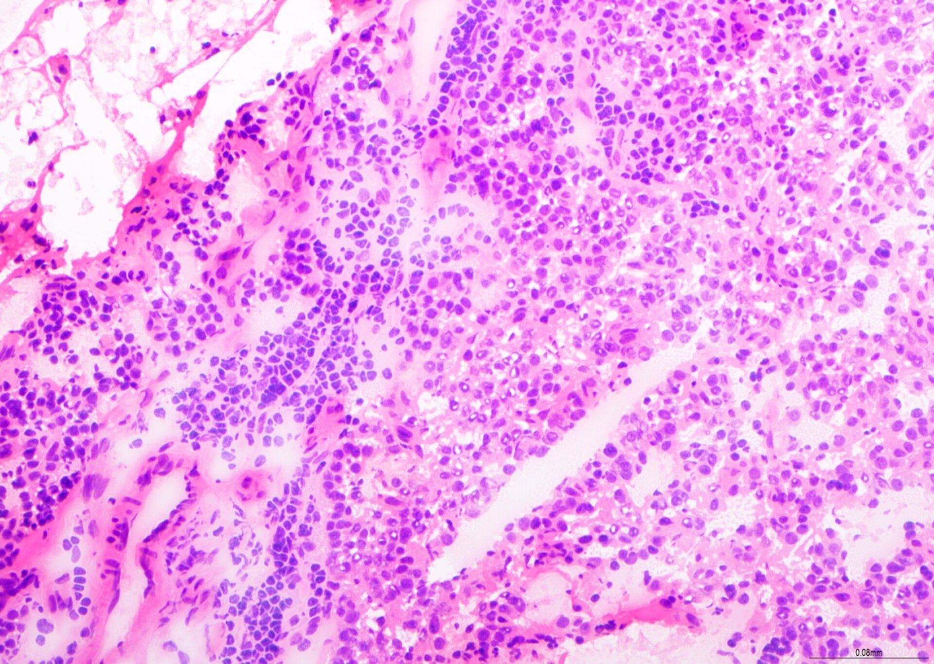
Haematoxylin & Eosin - Renal Scheme
Seen at x4, this is an example of a Haematoxylin & Eosin stained slide. In addition to the folds, creases and lifting, marks were also deducted for Haematoxylin background staining and weak eosin intensity.
This slide was scored 7/10.

Mohs Scheme
Seen at x4 magnification, heres a slide from the Mohs scheme. Marks were deducted for: folds and creases, knife debris, holes, trimming artefact, squames and floaters, eneven staining and water present. This slide scored 5/10 at assessment.
Haematoxylin & Eosin - Frozen Companion Scheme
Seen at x20, this is an example of an H+E. There us very little eosin staining which makes architectual assessment challenging. On that basis this section score a 5/10.
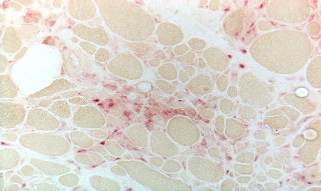
Acid Phosphatase - Muscle Scheme
Acid Phospahte can be demonstrated by hydrolysis. Staining conditions must be careully controlled to ensure specific acid phosphate demonstration, with an acidic pH found to be optimal. This is an excellent example where the preperation is free of deposit, and there is specific staining of acid phosphare activity againsta a clean background. This slide scored 9/10.
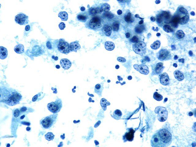
Head and Neck sample PAP - Non Gynae Scheme
This slide shows a mono layer of cells, no overlapping or clumping and excellent demonstration of chromatin detail. This slide scored 10/10 at assessment.
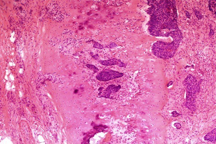
H+E - Mohs Scheme
Here's a H+E slide showing basal cell carcinmoa invasion into cartilage from tissue taken from the nasal bridge are. Marks were deducted for: Eosin intensity too string, knife marks, folds and creases. This slide scored 7/10 at assessment.